

The First Clinical Enrollment of the TriCap Tricuspid Valve Edge‑to‑Edge Repair Completed at the First Affiliated Hospital of Chongqing Medical University
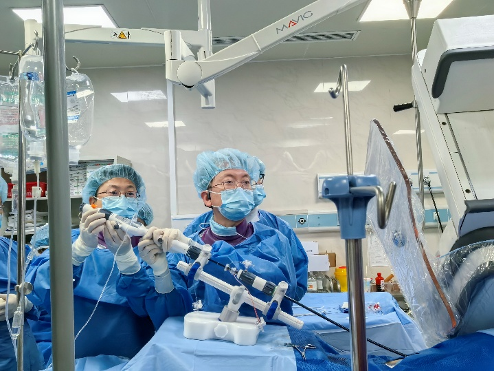
In the field of cardiac interventional therapy, the tricuspid valve was once called "the forgotten valve" due to its complex anatomy and irregular annulus. However, with the continuous breakthroughs of domestic innovative devices, this "forgotten corner" is now ushering in its high-light moment. Recently, a team led by Professor Luo Suxin, Professor Xiang Rui, Professor Yang Yuan, and Professor Dong Qian at the First Affiliated Hospital of Chongqing Medical University successfully performed, for the first time, a TriCap transcatheter tricuspid valve edge-to-edge repair procedure on a patient with quadricuspid valve malformation complicated by severe tricuspid regurgitation. After the procedure, the regurgitation was dramatically reduced from severe (4+) to mild (1+), "winning back" for the patient a long-missed comfortable life through a minimally invasive battle requiring "no thoracotomy and no cardiac arrest."
More than 2 years of exertional dyspnea, tricuspid regurgitation 4+ — a heart valve that "cannot close"
The patient is a 72-year-old female with a chief complaint of "exertional dyspnea for more than 2 years," diagnosed with valvular heart disease for more than 1 month. More than 1 month ago, after catching a cold, the patient's chest tightness and shortness of breath significantly worsened, and she was hospitalized at an outside hospital. During the hospitalization, coronary heart disease was diagnosed, and echocardiography suggested severe tricuspid regurgitation (4+). Symptomatic medical treatment including diuretics and inotropes was administered; after discharge, she continued oral medication for more than 1 month. The patient now presented to the First Affiliated Hospital of Chongqing Medical University for further diagnosis and treatment and was admitted through the outpatient clinic with a diagnosis of "severe tricuspid regurgitation."
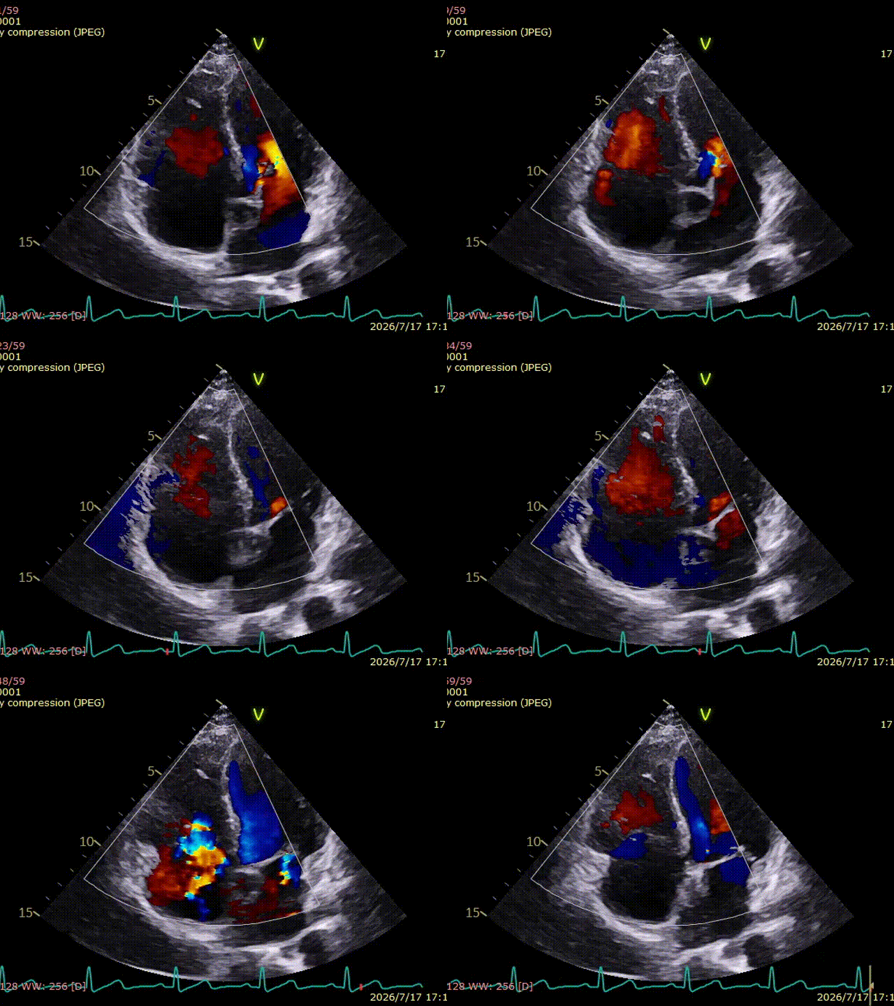
Preoperative transthoracic echocardiography assessment
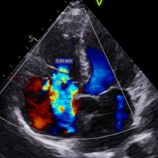
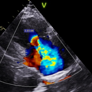
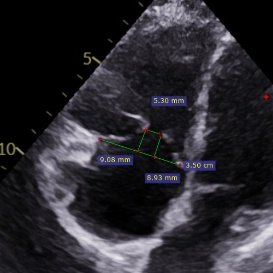
Severe tricuspid regurgitation (4+, mean vena contracta: 10 mm), with a predominantly central regurgitant orifice, combined with some near-central anteroseptal and posterior components. A 5.3 × 4.6 mm GAP was present centrally. Mild tethering of some leaflets (tethering height: 9 mm). Tricuspid annular dilatation (mean annular diameter: 41.5 mm). Pulmonary artery systolic pressure (PASP): 46 mmHg.
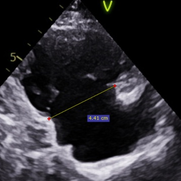
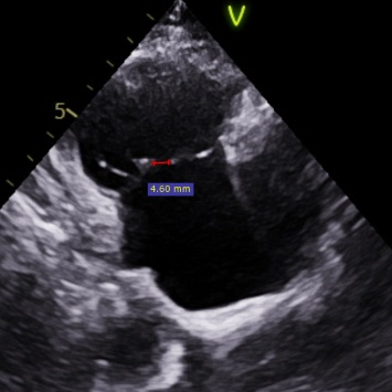
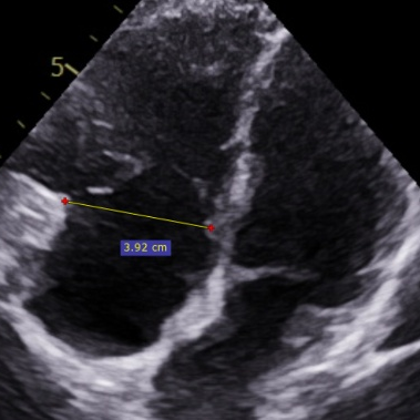
Type IIIa quadricuspid valve malformation + large GAP — how does TriCap "clamp" the hope of life?
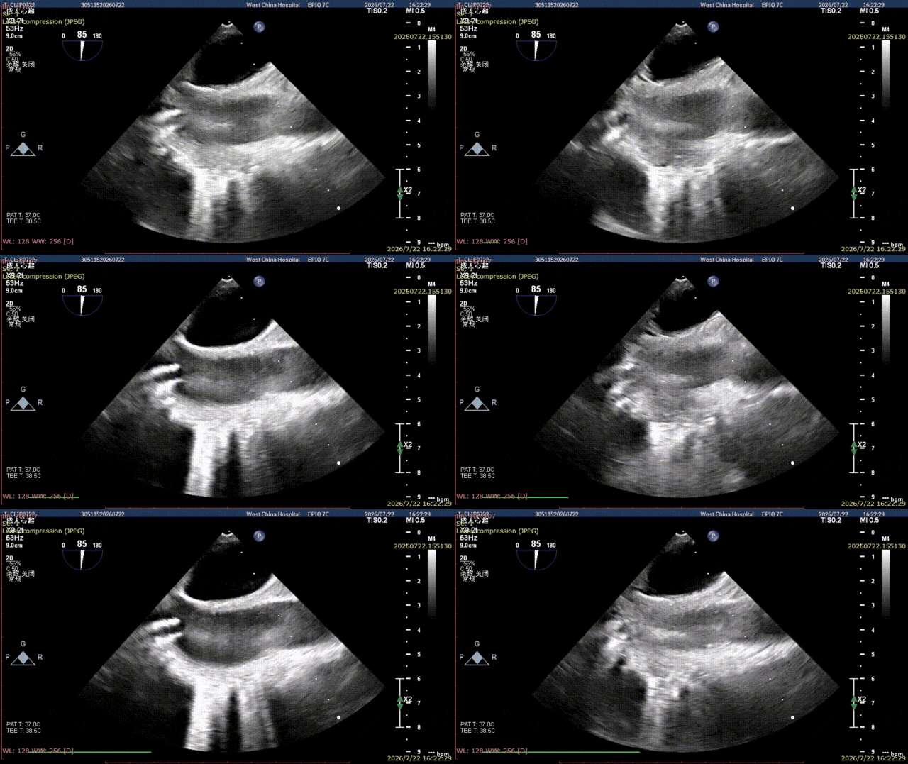
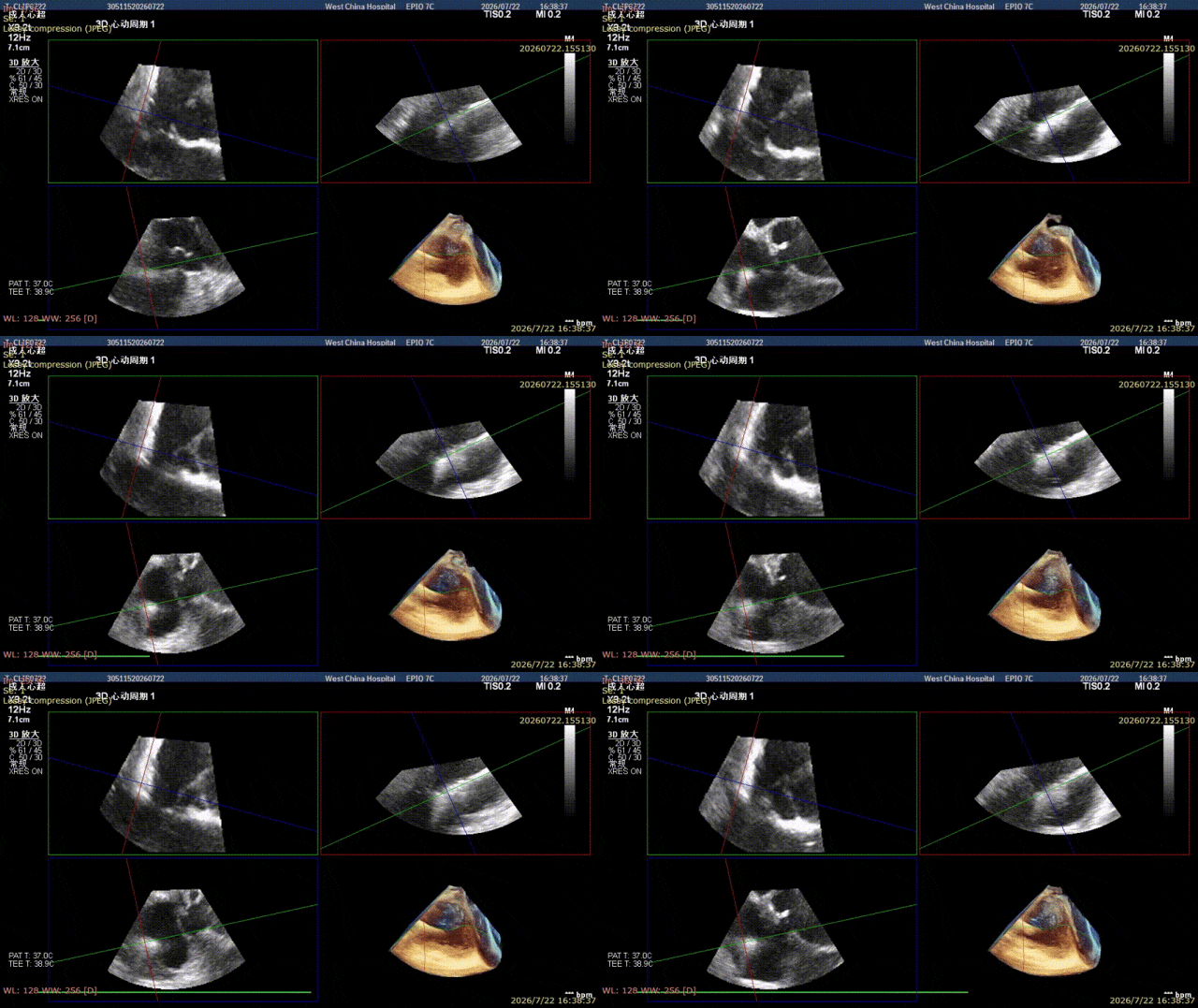
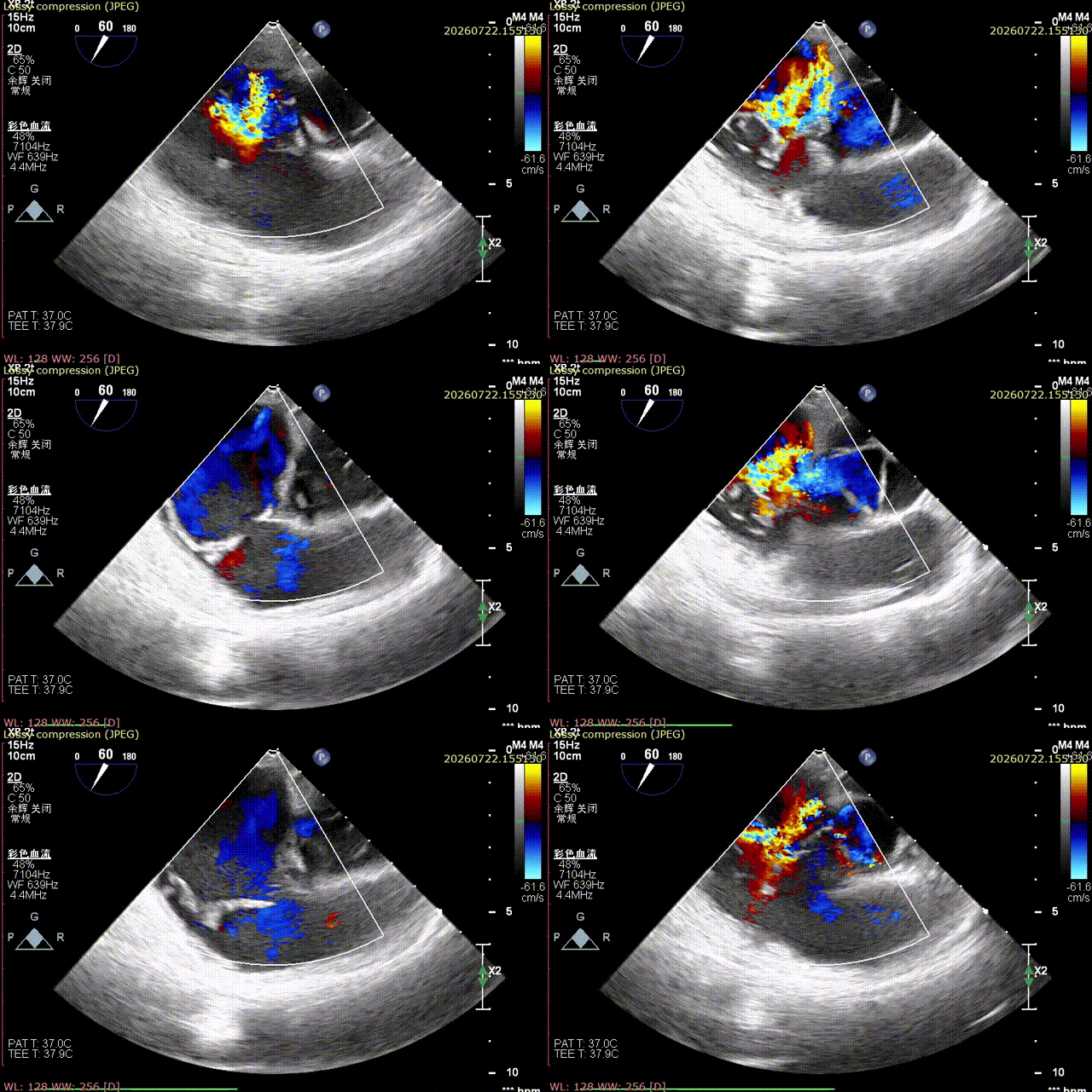
Transesophageal echocardiography revealed that the patient had a Type IIIa quadricuspid valve malformation: the anterior leaflet consisted of two leaflets (A1/A2).
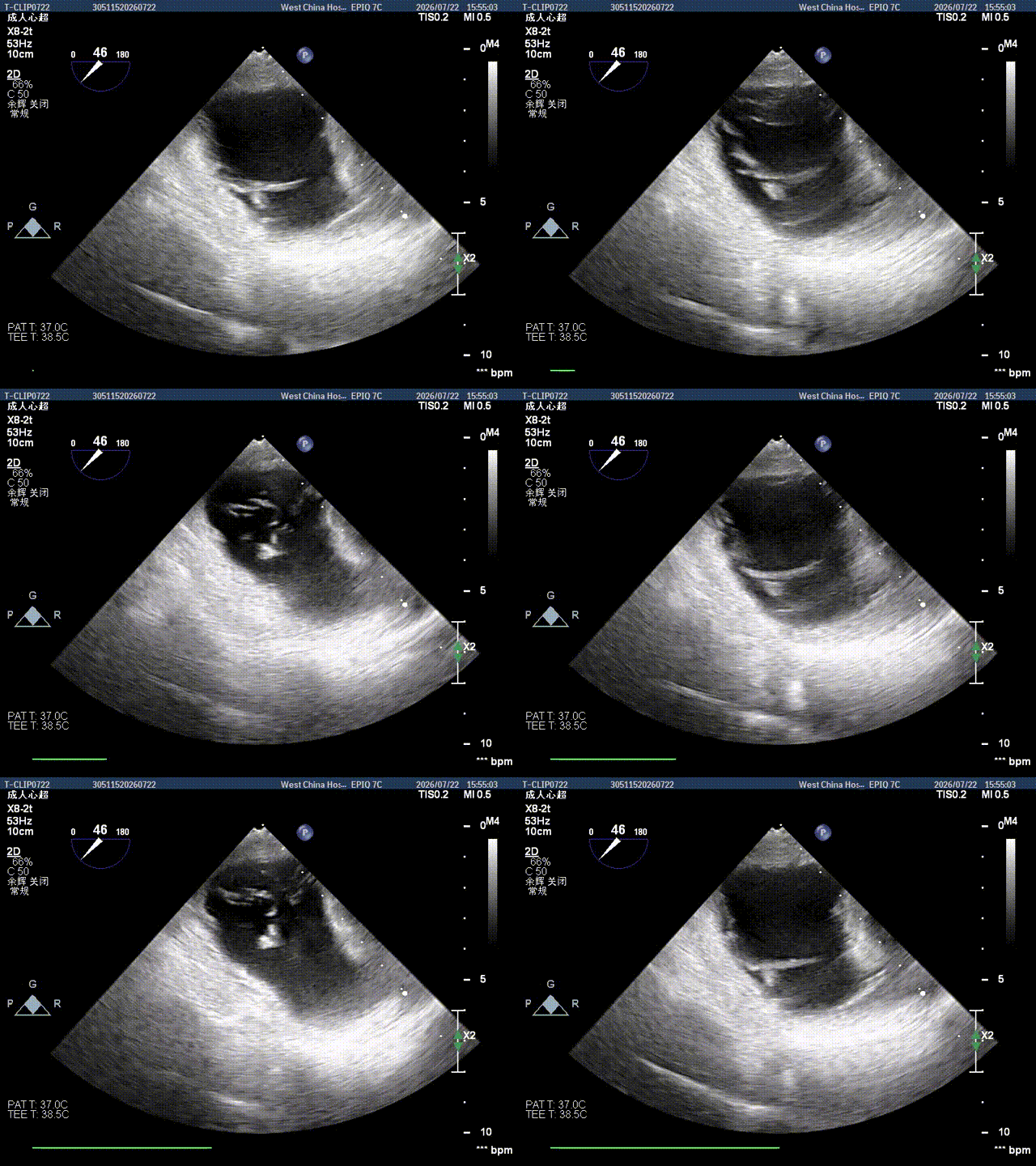
TEE transgastric baseline
After understanding the patient's condition, the cardiac team at the First Affiliated Hospital of Chongqing Medical University promptly conducted in-depth discussions and comprehensive evaluations together with the departments of ultrasound, anesthesiology, and imaging to formulate the surgical strategy. The plan was to use the TriCap transcatheter tricuspid valve edge-to-edge repair system to clip the A1/S and A2/S regions respectively, in order to improve the regurgitation.
Facing the triple challenge of Type IIIa quadricuspid valve malformation, severe annular dilatation, and a large coaptation gap, traditional open-heart surgery carries significant trauma and high risk. The TriCap transcatheter tricuspid valve edge-to-edge repair system is the first transcatheter tricuspid leaflet repair system in China with a "physiological tension" design. Compared with traditional rigid clipping devices, TriCap's clip arms can conform to the fragile tricuspid leaflet tissue, providing a flexible and well-apposed gripping force when capturing the leaflets, thus avoiding rigid tearing of the leaflet tissue.
Surgical procedure
After the patient received general anesthesia, the femoral vein was punctured intraoperatively to establish access. Relying on extensive experience and the flexible positioning capability of the TriCap steerable system, the operator precisely delivered the delivery system to the target region of the tricuspid valve under transesophageal echocardiography guidance alone.
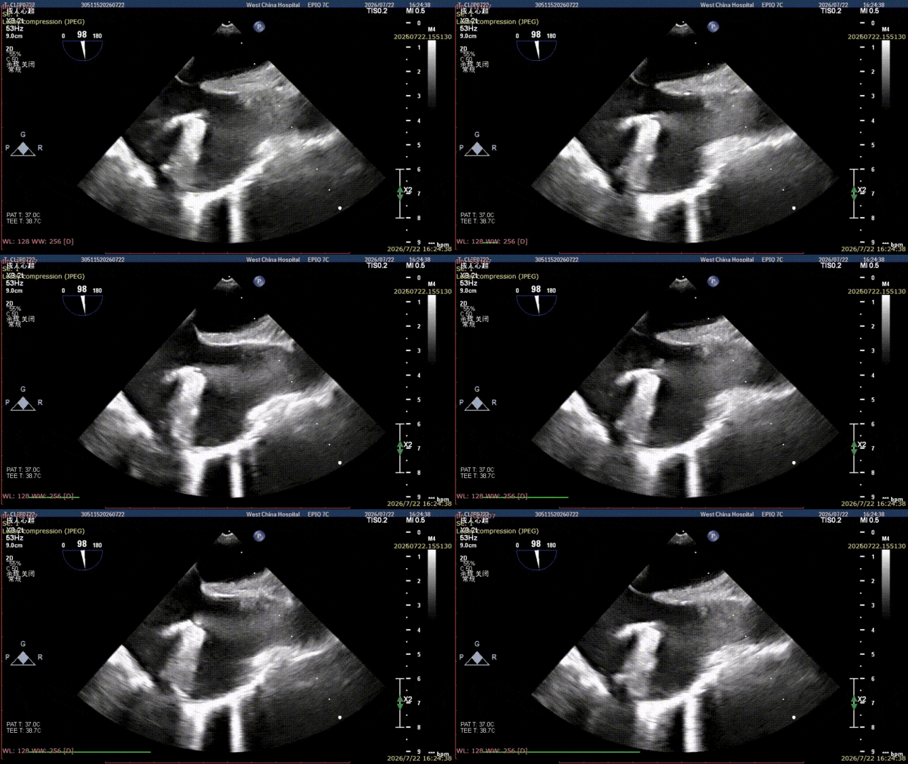
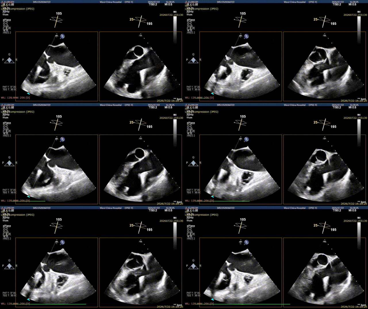
The device orientation was adjusted so that the device was aligned with the A1-S commissural region of the tricuspid valve. The clipping device was advanced across the tricuspid orifice into the right ventricular side, and then slowly withdrawn.
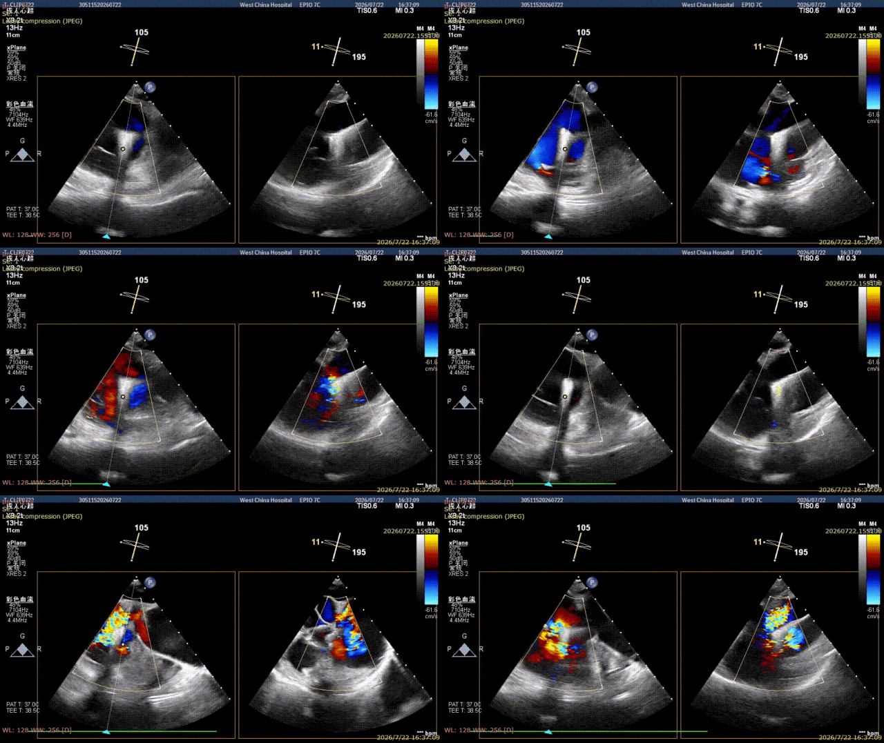
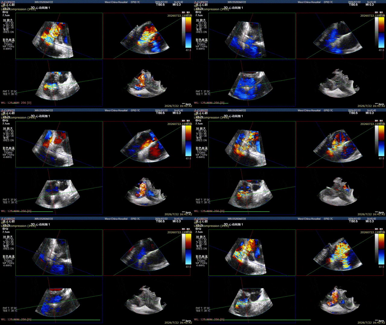
Under ultrasound confirmation, the leaflets were captured. After confirming sufficient and stable grasping of the leaflets, the clip was closed; immediate assessment showed that the A1-S regurgitation had almost disappeared, with only residual central regurgitation. The first clip was then released.
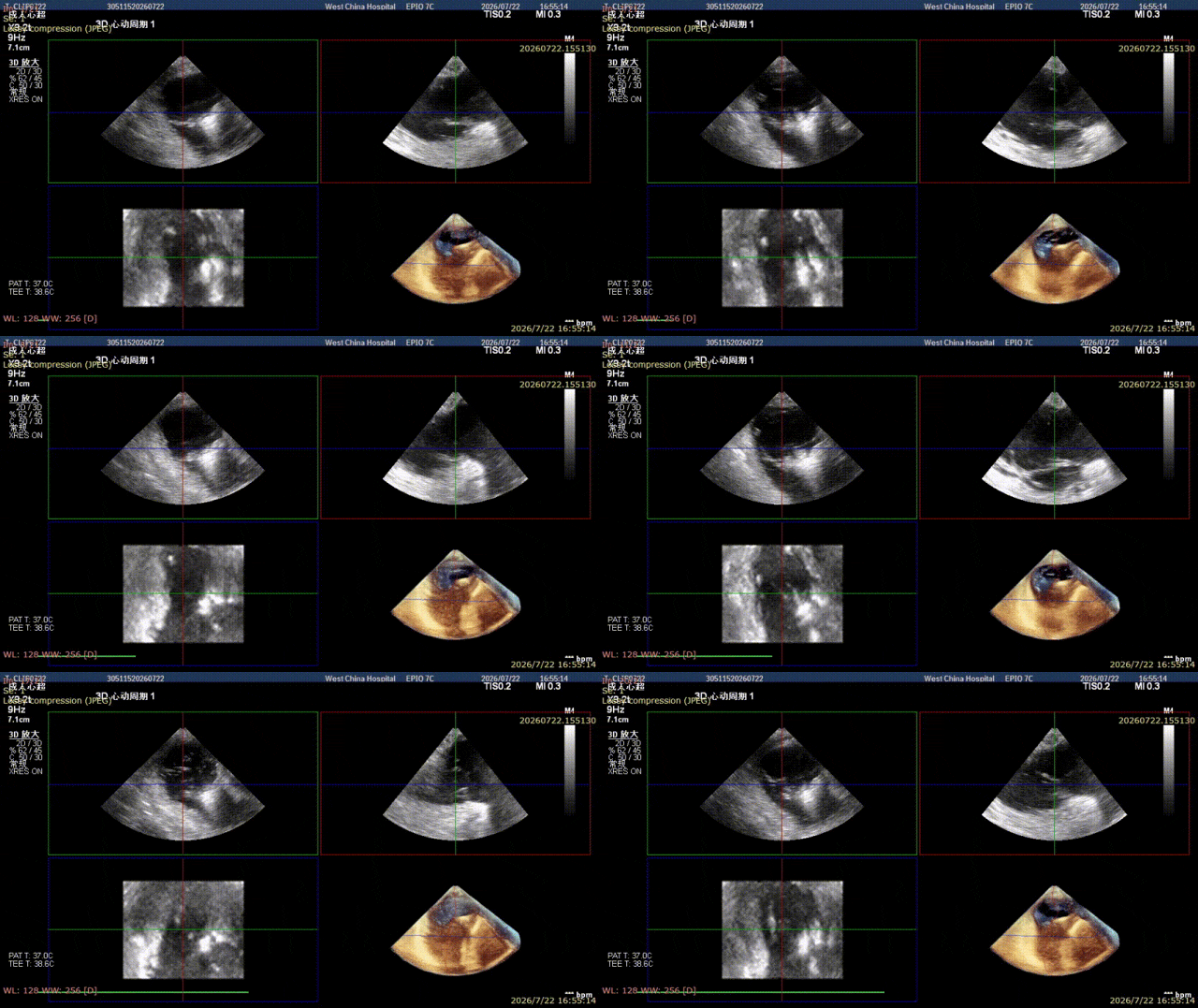
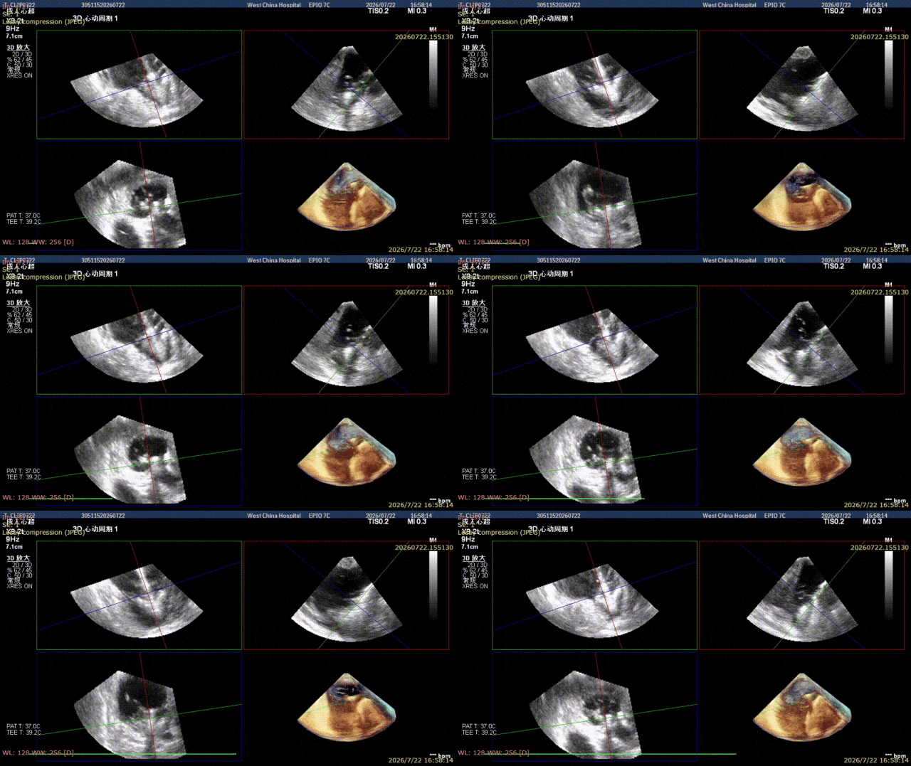
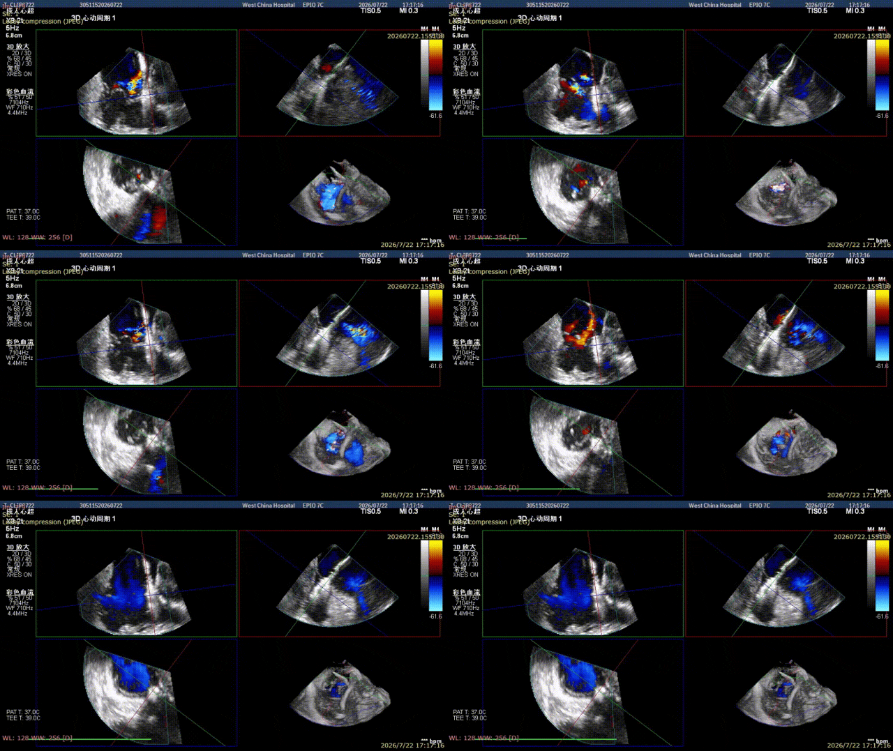
Subsequently, a second clip was delivered along the delivery system, aimed at the A2-S region near the center. During the first grasping attempt, it was found that the clip was located at the P-S commissure, resulting in less capture of the anterior leaflet. The clip was opened and its axis was slightly adjusted to be closer to A2-S. After the clip was closed, only trace regurgitation remained. Transthoracic echocardiography indeed showed tricuspid regurgitation of 1+ (trace).
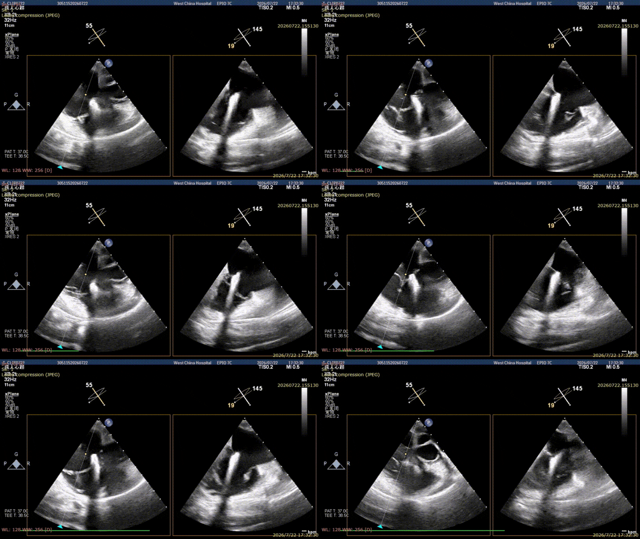
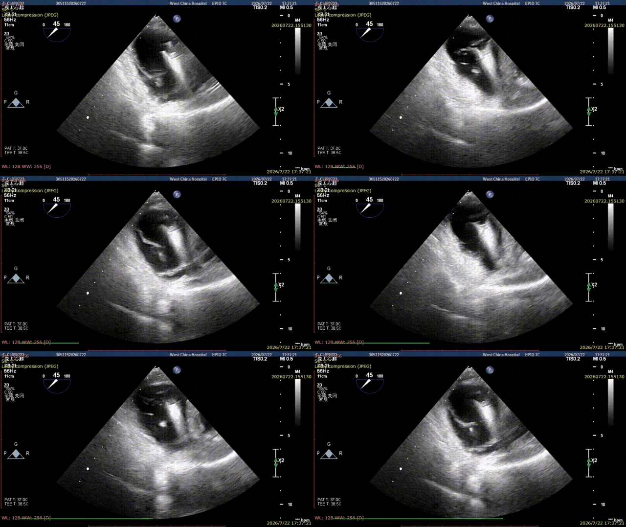
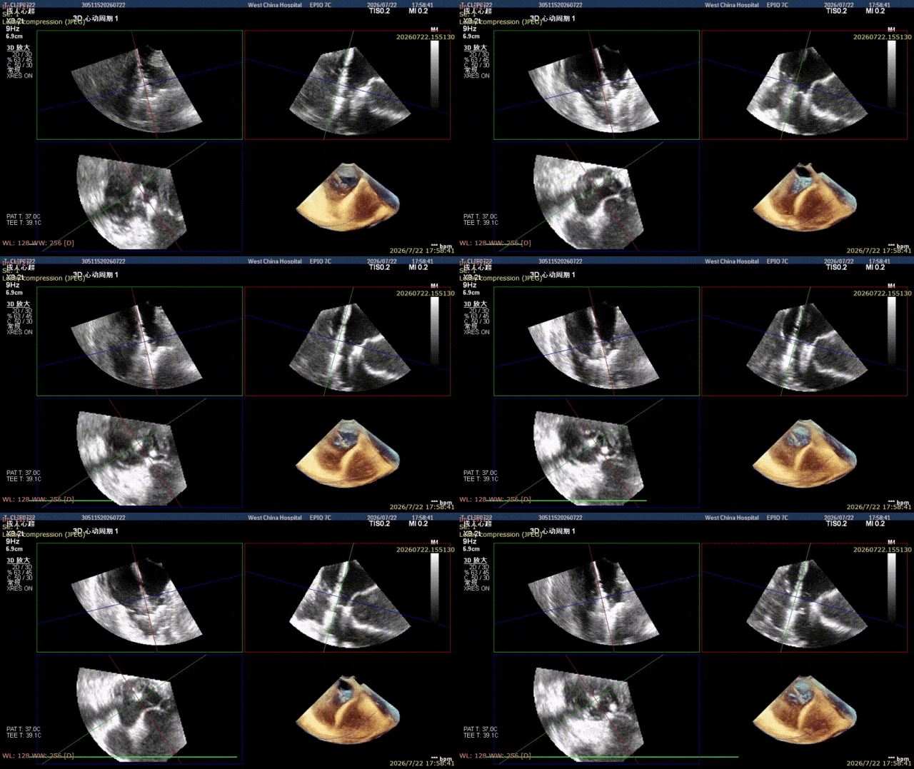
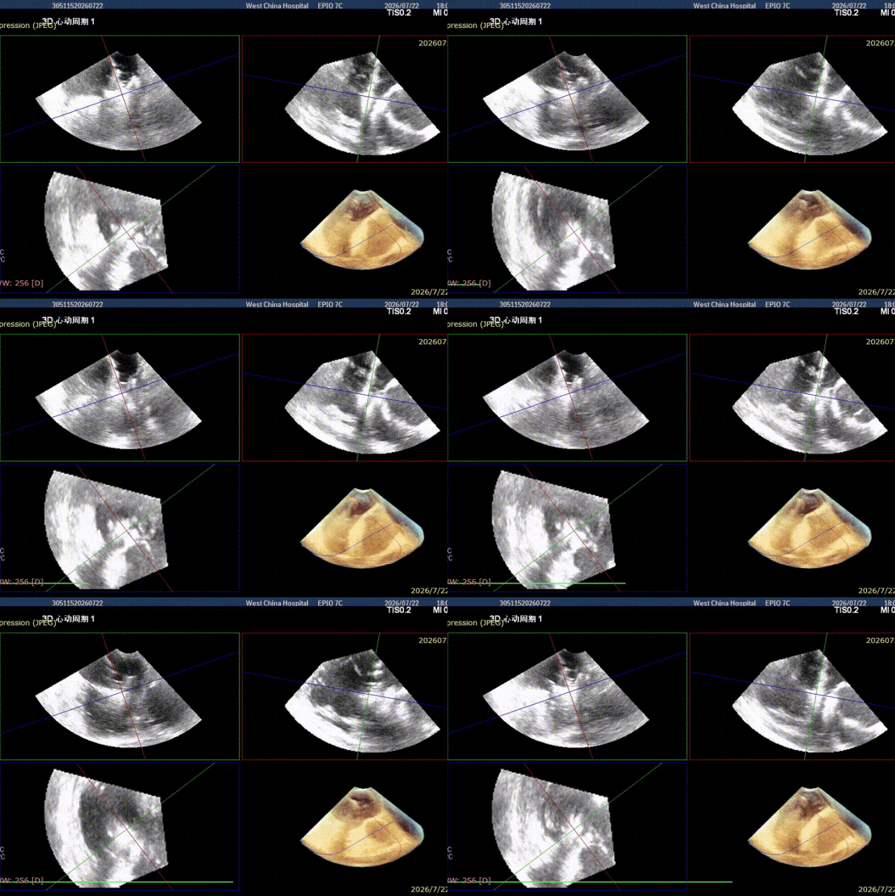
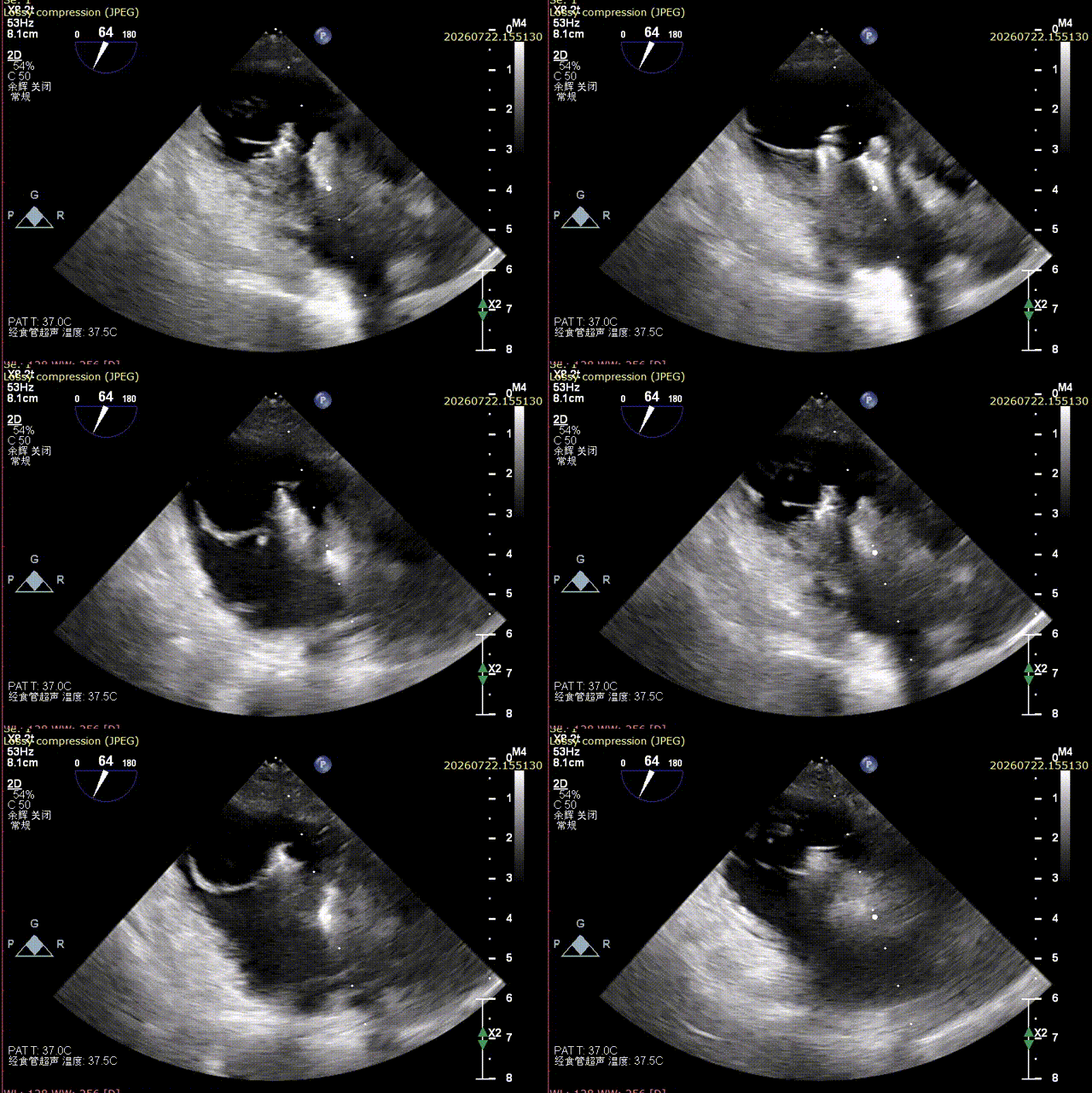
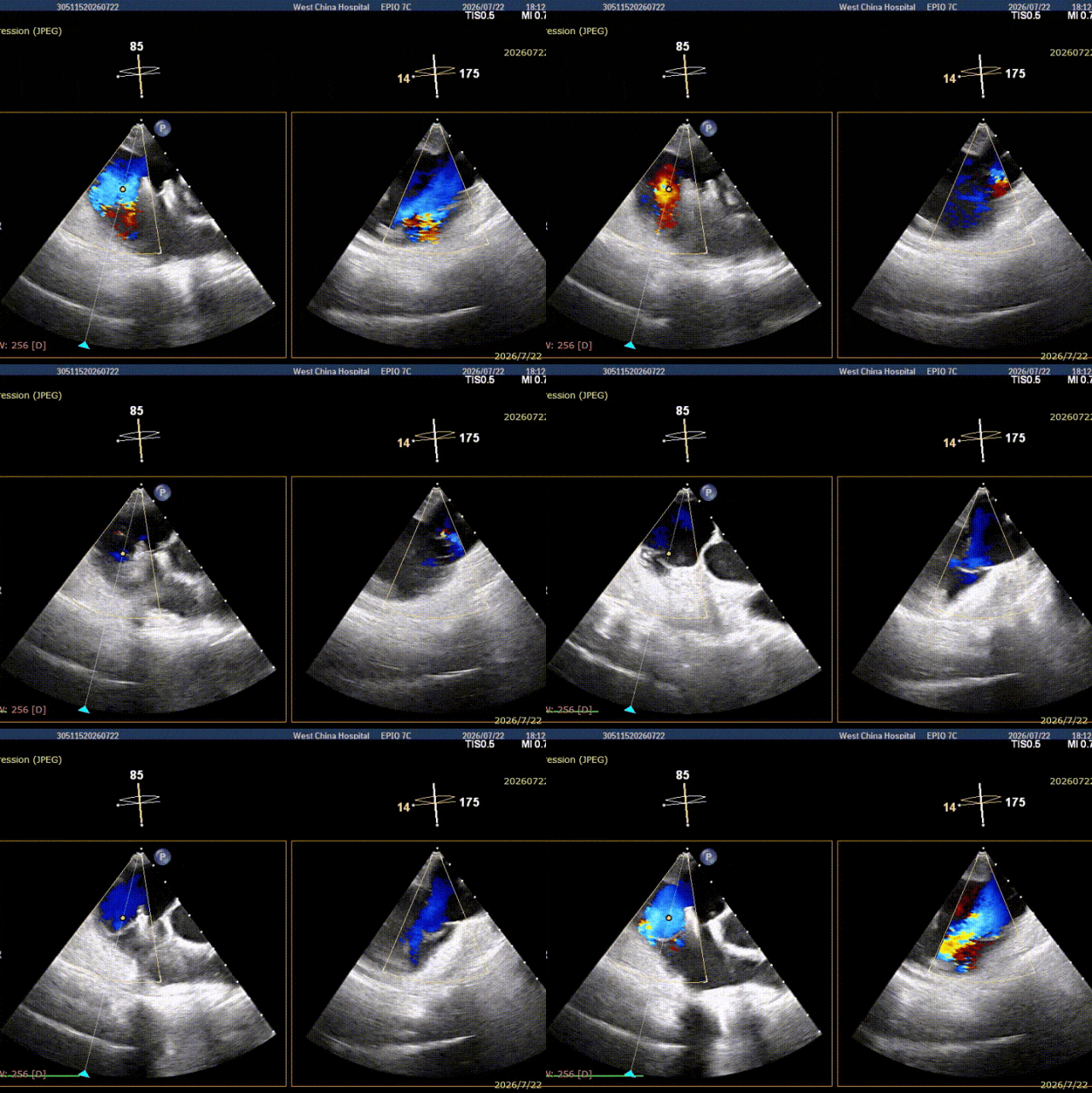
Tricuspid annular peak transvalvular pressure gradient: 4 mmHg; mean transvalvular pressure gradient: 2 mmHg.
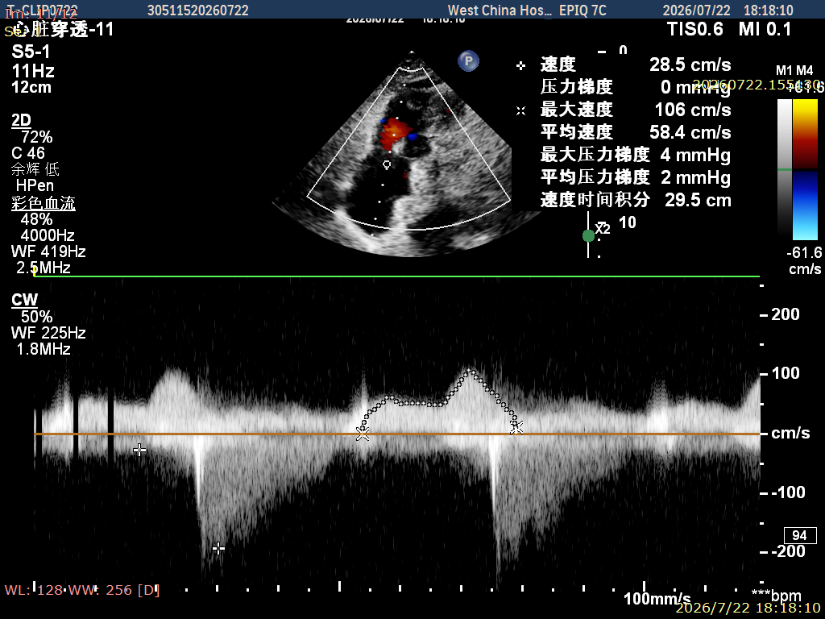
Two clips, two precise "captures" — the transformation from 4+ to 1+
From severe regurgitation of 4+ before the procedure to mild residual regurgitation of 1+ immediately after the procedure, the surgical team completed this "valve defense battle" with two clean and decisive leaflet clips. Intraoperatively, the two TriCap clips precisely captured the leaflets in the A1/S and A2/S regions respectively, successfully closing the coaptation gap. Immediate postoperative ultrasound showed a significant reduction in the degree of regurgitation and a marked improvement in hemodynamics.
Preoperative vs. postoperative regurgitation comparison images:
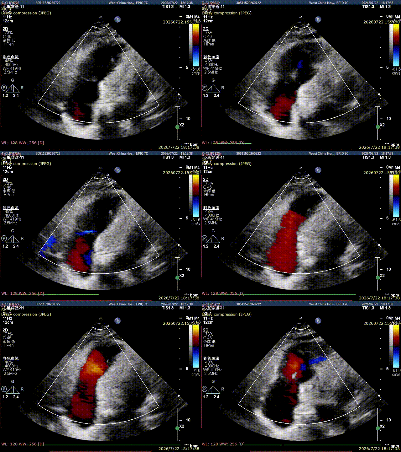
TTE
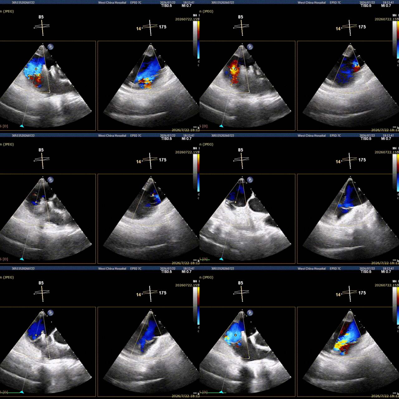
TEE
Conclusion

The tricuspid valve — this once "forgotten valve" — is being seen anew thanks to the power of domestic innovation. With its three major advantages of physiological tension, flexible gripping, and minimally invasive access, the TriCap transcatheter tricuspid valve edge-to-edge repair system provides patients with severe tricuspid regurgitation a new option that is safe, efficient, and minimally traumatic. The success of this procedure not only validates the outstanding performance of domestic innovative devices in the face of complex tricuspid valve pathologies — including quadricuspid valve malformation, large coaptation gaps, and severe annular dilatation, these "hard bones to chew" — but also demonstrates the profound expertise and cutting-edge vision of Professor Xiang Rui's cardiac team at the First Affiliated Hospital of Chongqing Medical University in the field of structural heart disease interventional therapy. From precise preoperative assessment to meticulous surgical strategy, from refined intraoperative maneuvers to seamless multidisciplinary collaboration, every step reflects patient-centered medical warmth and the courage to be a pioneer with technical confidence. From 4+ to 1+, what changed is the number; what remains unchanged is the medical profession's solemn commitment to every heart. As domestic innovative technologies such as TriCap continue to be promoted, more patients with complex tricuspid valve pathologies will have the opportunity to avoid the suffering of thoracotomy and regain "heartfelt" new life in the minimally invasive era.
Expert profiles


Xiang Rui
The First Affiliated Hospital of Chongqing Medical University

Yang Yuan
The First Affiliated Hospital of Chongqing Medical University

Dong Qian
The First Affiliated Hospital of Chongqing Medical University


